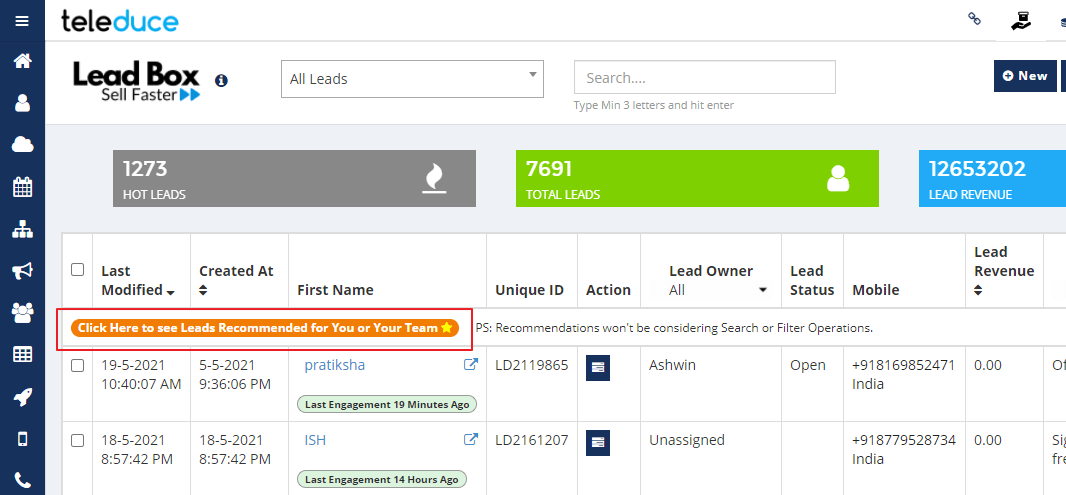
Glymphatic System Key to Parkinson’s Protein Spread and Aggregation

Summary: Researchers shed light on the puzzle of Parkinson’s disease progression. They discovered that a mutated version of the α-synuclein protein moves through the brain’s lymphatic system, known as the glymphatic system, before aggregating.

By tracking fluorescent α-synuclein in mice, they observed the protein’s early spread, with fibril formation much later. This suggests that addressing the monomeric α-synuclein and its propagation may halt Parkinson’s progression.

Key Facts:

  1. The mutated α-synuclein protein propagates through the glymphatic system before forming clumps.
  2. Fluorescent α-synuclein appeared in distant brain areas just two weeks post-injection, showing early propagation.
  3. Fibrils of α-synuclein only formed 12 months post-injection, highlighting the delay between propagation and aggregation.

Source: Tokyo Medical and Dental University

In many neurodegenerative disorders, abnormal proteins progressively aggregate and propagate in the brain. But what comes first, aggregation or propagation? Researchers from Japan share some new insights about the mechanism involved in Parkinson’s disease.

In a study published recently in Cell Reports, researchers from Tokyo Medical and Dental University (TMDU) have shown that a mutated version of a protein called α-synuclein propagates to various cerebral regions through the lymphatic system and then aggregates.

This shows neurons.
Interestingly, fluorescent α-synuclein was detected in remote regions two weeks after injection, indicating an early spreading of mutant α-synuclein in the brain. Credit: Neuroscience News

Although the function of α-synuclein is not fully understood, it participates in neurotransmission. However, in some neurodegenerative diseases including Parkinson’s disease, α-synuclein changes shape and forms pathological clumps.

“Most experiments conducted so far only used fibrils, which are the clumps formed when monomeric α-synuclein aggregates. The fibrils are transmitted from neurons to neurons, but it remains unclear whether monomers act in the same way,” explains Kyota Fujita, an author of the study.

To further investigate how monomers and fibrils of α-synuclein move around in the brain, the researchers injected small amounts of viral particles into the orbital cortex of mice to produce fluorescent monomeric mutant α-synuclein.

Because any cell type can contribute to α-synuclein propagation, they used viral particles to enable the synthesis of α-synuclein monomers in all cell types present in the injection area. This method ensured that all modes of propagation were accounted for.

Twelve months after the injection, although the fluorescent signal was lower in the injected region, signals were detected in other brain areas. Interestingly, fluorescent α-synuclein was detected in remote regions two weeks after injection, indicating an early spreading of mutant α-synuclein in the brain.

But how did α-synuclein propagate? The team followed the three-dimensional distribution of α-synuclein in the brain and found fluorescent α-synuclein in the glymphatic system, which is the lymphatic system of the brain.

The glymphatic system is involved in draining and renewing fluid from the brain and eliminating toxins, but it could also distribute toxic substances throughout the brain. The team also observed the presence of fluorescent α-synuclein in the matrix surrounding neurons and in the cytosol of neurons.

This finding suggested that fluorescent α-synuclein was taken up by the extracellular matrix and, subsequently, by neurons.

The researchers also investigated the aggregation state of α-synuclein in the remote brain regions. “Fibrils of α-synuclein formed after the monomers had propagated,” says Professor Hitoshi Okazawa, the research group leader.

“Specifically, we observed α-synuclein monomer in the glymphatic system and remote regions as early as two weeks after injection, while we found α-synuclein fibrils 12 months after injection!”

The amount of α-synuclein aggregated and the time at which they formed after injection varied among regions and was not proportional to the distance from the injection site. This observation is consistent with the known vulnerability of some regions to pathological α-synuclein.

This study shows how monomeric α-synuclein propagates through the glymphatic system in a different way from the fibrils. Thus, targeting these early events, α-synuclein monomer and  brain lymphatic system,  may limit the progression of Parkinson’s disease.

About this Parkinson’s disease research news

Author: Hitoshi Okazawa
Source: Tokyo Medical and Dental University
Contact: Hitoshi Okazawa – Tokyo Medical and Dental University
Image: The image is credited to Neuroscience News

Original Research: Open access.
Mutant α-synuclein propagates via the lymphatic system of the brain in the monomeric state” by Hitoshi Okazawa et al. Cell Reports


Abstract

Mutant α-synuclein propagates via the lymphatic system of the brain in the monomeric state

Highlights

  • Prionoid protein propagation is a common mechanism of neurodegenerative diseases
  • Neuron-to-neuron propagation of aggregated disease proteins has been assumed
  • We reveal propagation of the monomer disease protein via brain lymphatic system
  • The brain lymphatic propagation of monomer is a target of future therapeutics

Summary

Prion-like protein propagation is considered a common pathogenic mechanism in neurodegenerative diseases.

Here we investigate the in vivo propagation pattern and aggregation state of mutant α-synuclein by injecting adeno-associated viral (AAV)-α-synuclein-A53T-EGFP into the mouse olfactory cortex.

Comparison of aggregation states in various brain regions at multiple time points after injection using western blot analyses shows that the monomeric state of the mutant/misfolded protein propagates to remote brain regions by 2 weeks and that the propagated proteins aggregate in situ after being incorporated into neurons.

Moreover, injection of Alexa 488-labeled α-synuclein-A53T confirms the monomeric propagation at 2 weeks. Super-resolution microscopy shows that both α-synuclein-A53T proteins propagate via the lymphatic system, penetrate perineuronal nets, and reach the surface of neurons.

Electron microscopy shows that the propagated mutant/misfolded monomer forms fibrils characteristic of Parkinson’s disease after its incorporation into neurons.

These findings suggest a mode of propagation different from that of aggregate-dependent propagation.

Join our Newsletter
I agree to have my personal information transferred to AWeber for Neuroscience Newsletter ( more information )
Sign up to receive our recent neuroscience headlines and summaries sent to your email once a day, totally free.
We hate spam and only use your email to contact you about newsletters. You can cancel your subscription any time.